
Every scan we perform
The complete list, straight from our own catalogue. Search it or filter it — 267 studies across seven services, all under one roof in Bajajnagar.
Brand-new 3T scanner, first in Bajajnagar and Waluj
Brain
About this group▾
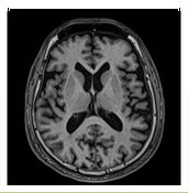 Actual image from Veda
A detailed scan of the brain using a magnetic field — no X-rays at all. Shows stroke, tumour, infection, multiple sclerosis, and the cause of headache, fits or weakness.
Nothing special. Eat and drink normally and take your usual medicines. Remove all metal — jewellery, watch, hairpins, hearing aid, dentures. Tell us if you have a pacemaker, cochlear implant, aneurysm clip or any metal inside your body.
20 – 25 minutes. You must lie still. The machine is loud and we give you earplugs.
Same day. Urgent cases within 2 hours.
- MRI Brain — plain
- Brain + Angiography (MRA)
- Brain + Venography (MRV)
- MR Stroke protocol
- Brain epilepsy protocol
- Brain + Orbit
- MR Pituitary
- Functional imaging (fMRI)
- Tractography
- MR Spectroscopy
- MR Perfusion — tumour grading and recurrence
- SWI — microbleeds and diffuse axonal injury
Spine
About this group▾
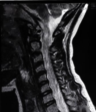 Actual image from Veda
Images the spinal bones, the discs between them and the spinal cord itself. The usual study for back or neck pain, sciatica, numbness and weakness in the arms or legs.
No fasting. Empty your pockets and remove belts and metal items. Tell us if you have had spine surgery or have any implant, and bring the operation notes if you can.
15 – 20 minutes for one region. A whole-spine screen takes about 30 minutes.
Same day.
- Cervical spine
- Thoracic spine
- Lumbo-sacral spine
- W.S.S — whole spine screening
Head & neck
About this group▾
Covers the ear, the jaw joint, the salivary glands, the mouth and throat. Used for hearing loss, ringing in the ear, dizziness, jaw pain and clicking, a lump in the neck, and staging of head and neck cancers.
No fasting for a plain study. Remove earrings, nose pins, chains and any removable denture. For the jaw joint we scan with your mouth open and closed, so tell us if opening your mouth is painful.
20 – 30 minutes.
Same day.
- Temporal bone
- Oral cavity + oropharynx
- Salivary gland tumours
- Other head & neck cancers
- MRI Temporomandibular joint (TMJ)
- MRI Orbit
- MRI Internal auditory canal / CP angle
- MR Sialography
Chest
About this group▾
Looks at the space between the lungs, the thymus, the chest wall — and the heart itself. Cardiac MRI measures how well the heart muscle is pumping and whether any of it has been damaged.
For a cardiac study come after 4 hours of fasting, bring your ECG and echo reports, and avoid tea, coffee and any stimulant that morning. A plain chest MRI needs no preparation.
Cardiac MRI takes 40 – 50 minutes with repeated breath-holds. A plain chest study takes 20 minutes.
Cardiac studies the same day, discussed with your cardiologist.
- Mediastinum — thymus
- MR Chest
- Cardiac MRI — function, viability and myocarditis
- MR Thoracic outlet syndrome
Breast
About this group▾
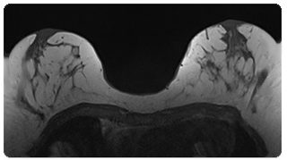 Actual image from Veda
The most sensitive breast study there is. Used to work out how far a known cancer extends, to screen women at high risk, and to answer a question the mammogram and ultrasound could not settle.
If you still have periods, book between day 7 and day 14 of your cycle. Contrast is given, so come after 4 hours of fasting with a recent creatinine report. Bring all previous mammograms and ultrasound films. You lie face down for this scan.
30 – 40 minutes.
Same day, with a BI-RADS category and a clear next step.
- Breast MRI — diagnosis, staging and BI-RADS
Abdomen
About this group▾
Liver, pancreas, kidneys, bowel and the bile ducts. MRCP maps the bile and pancreatic ducts without any injection or endoscopy; MR enterography shows the small bowel in Crohn's disease.
Nothing to eat or drink for 4 to 6 hours before. Bring any previous ultrasound or CT films. For enterography you will be asked to drink a special liquid in the hour before the scan.
20 – 30 minutes. You will be asked to hold your breath several times.
Same day.
- Abdomen plain + contrast
- MRCP — cholangiopancreatography
- MR Prostate
- MR Rectum
- MR Fistulogram
- MR Defecogram
- MR Renal / IVP
- MR Enterography — small bowel and Crohn's
- MR Prostate — multiparametric, PI-RADS v2.1
Pelvis
About this group▾
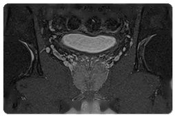 Actual image from Veda
The uterus, ovaries, prostate and rectum. Used for fibroids, endometriosis, ovarian cysts, staging of cancers of the womb or cervix, and multiparametric prostate MRI reported to PI-RADS.
Come after 4 hours of fasting with a moderately full bladder. For a prostate study, avoid ejaculation for 3 days before and tell us the date of any recent biopsy — we prefer to wait 6 weeks after one.
30 – 40 minutes for a prostate study, 25 minutes otherwise.
Same day.
- Endometrial / cervical / ovarian malignancy protocol
- Any pelvic tumour or cyst
- Congenital uterine anomalies
Musculoskeletal
About this group▾
 Actual image from Veda
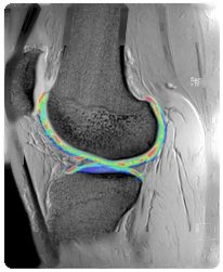
Joints, ligaments, cartilage, tendons and muscle. The standard study for a torn meniscus or cruciate ligament, a rotator cuff tear, hip impingement and unexplained joint pain. Cartilage mapping shows early wear before it is visible any other way.
No fasting. Wear loose clothes without metal. Tell us about any plate, screw, nail or joint replacement — most are safe, but we need to know which one and where.
15 – 20 minutes for one joint.
Same day.
- Shoulder
- Elbow
- Wrist / finger
- Hip
- Knee
- Ankle
- Mid-forefoot
- Sacroiliac joint
- Cartilage T2 mapping
- Hip — FAI and labral protocol
Whole body
About this group▾
 Actual image from Veda
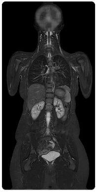
Head to thigh in one sitting, without a single X-ray. Used to search the whole skeleton and organs for spread of cancer, and in myeloma. Whole-body diffusion picks up deposits that a bone scan can miss.
Come after 4 hours of fasting. Bring every previous scan and report you have — this study is most useful when it can be compared. Allow a longer appointment.
45 – 60 minutes.
Same day.
- Whole body MRI
- Whole body MR Angiography
- Whole body diffusion (DWIBS) — myeloma and metastatic screening
Nerve & plexus
About this group▾
MR neurography images the nerves themselves — the brachial plexus in the shoulder, the lumbosacral plexus, and peripheral nerves. For shoulder or arm pain after injury, unexplained numbness, and nerve tumours. A 3T scanner is what makes this possible.
No fasting. Bring your nerve conduction study if you have had one — it tells us exactly which nerve to concentrate on.
30 – 40 minutes.
Same day.
- MR Neurography — peripheral nerve
- Brachial plexus
Angiography
About this group▾
 Actual image from Veda
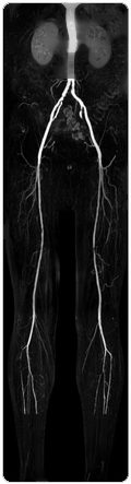
Maps arteries and veins without a catheter and without X-rays. Brain and neck vessels, the aorta, the leg run-off, and limb or pelvic veins for suspected clot.
Some studies need a contrast injection — for those, come after 4 hours of fasting with a recent creatinine report. Brain and neck angiography usually needs no injection at all.
25 – 35 minutes.
Same day.
- MR Aortogram and peripheral run-off
- MR Venography — limb and pelvic
Fetal MRI
About this group▾
An MRI of the baby while still in the womb, used when the ultrasound has found something in the brain or spine that needs a closer look. There is no radiation of any kind.
Nothing to eat for 4 hours. Bring the referring doctor's note and your anomaly scan report. Best done after 20 weeks.
25 – 30 minutes, lying on your back or on your side.
Same day, discussed directly with your obstetrician.
- Fetal MRI — brain and spine
- MR Placenta — accreta spectrum
Children
About this group▾
Fast MRI protocols built for children, so that most scans can be done without sedation. A parent stays in the room throughout.
Bring a familiar toy or blanket. For an infant, time the appointment just after a feed so the baby sleeps through. Tell us in advance if sedation has been advised, so that we can arrange it properly.
10 – 20 minutes for most fast protocols.
Same day.
All plain scans, all contrast scans, all angiographies
Brain
About this group▾
 Actual image from Veda
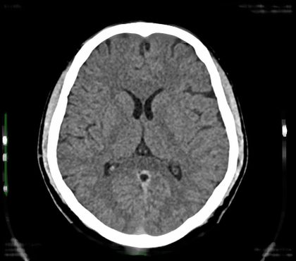
The fastest way to look inside the head. In a stroke, a head injury or a sudden severe headache it shows bleeding, swelling and fracture within seconds of the scan finishing.
Nothing at all for a plain scan — you can walk in at any hour. Remove earrings, hairpins and spectacles. If contrast is needed, come after 4 hours of fasting with a recent creatinine report.
Under a minute inside the scanner. About 10 minutes in the room altogether.
Emergencies within the hour, at any time of night. Routine studies the same day.
- CT Brain plain
- CT Brain contrast / venography
PNS & face
About this group▾
 Actual image from Veda
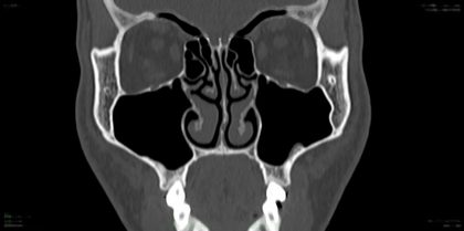
Thin slices through the sinuses, the facial bones, the eye socket and the temporal bone. For long-standing blocked nose and sinusitis before surgery, facial fractures, and hearing loss or discharge from the ear.
None for a plain study. Remove spectacles, earrings, nose pins and any removable denture. Bring your ENT surgeon's note — it tells us how fine a slice to take.
Under a minute.
Same day, usually within 2 hours.
- CT PNS plain
- CT PNS contrast
- Face 3D (orbit / mandible / TMJ)
- HRCT Temporal bone / inner ear
- CT Orbit
Thorax
About this group▾
 Actual image from Veda
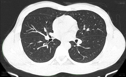
A high-detail CT of the lungs taken in very thin slices. For a long-standing cough, breathlessness, suspected fibrosis or tuberculosis, and the after-effects of infection. Low-dose screening is available for long-term smokers.
None. A plain HRCT needs no injection. Wear clothes without metal buttons or zips, and remove any chain. You will be asked to take a deep breath and hold it.
Under a minute inside the scanner; about 10 minutes in the room altogether.
Same day, usually within 2 hours.
- HRCT Thorax (chest)
- CT Thorax + contrast
- Low-dose lung cancer screening
Neck
About this group▾
Images the thyroid, the salivary glands, the throat and the lymph nodes of the neck. Used for a neck lump, difficulty swallowing, a deep abscess, and staging of head and neck cancer.
Contrast is usually needed, so come after 4 hours of fasting with a recent creatinine report. Remove chains and any removable denture.
About 10 minutes in the room.
Same day.
- CT Neck
- CT Neck + contrast
Joints — 3D
About this group▾
 Actual image from Veda
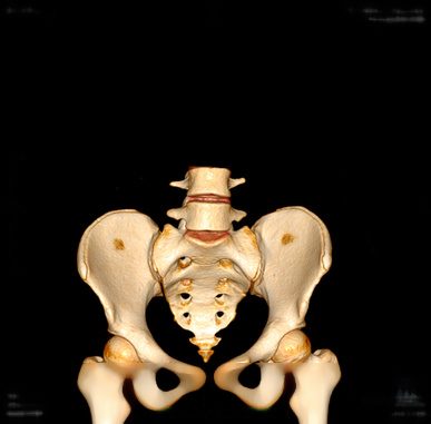
The bone rebuilt in three dimensions, so a surgeon can turn it on screen and see exactly how a fracture lies before operating. We can also supply the data as a 3D-printable file.
No preparation. Tell us if the limb is in a plaster cast — we can still scan through it. Bring the plain X-Ray if one has already been taken.
Under a minute to scan; the 3D rebuilding takes another 15 minutes.
Same day, with films and a CD for your surgeon.
- 3D Shoulder joint
- 3D Elbow joint
- 3D Wrist joint
- 3D Hip joint
- 3D Knee joint
- 3D Ankle joint
- 3D Foot joint
- STL export for 3D printing and surgical planning
Angiography
About this group▾
 Actual image from Veda
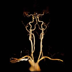
A dye injection with the scan running, so every artery fills and is mapped. Used for blocked leg arteries, aneurysm, suspected clot in the lung, and narrowing of the neck vessels before a stroke happens.
Come after 4 hours of fasting with a recent serum creatinine report. Tell us about any allergy to iodine or contrast, asthma, kidney disease, or if you take metformin — it usually has to be stopped for two days afterwards, on your doctor's advice.
About 15 minutes in the room, plus 20 minutes afterwards for the vessel reconstruction.
Same day. Emergencies within the hour.
- Brain angiography
- Neck and carotid angiography
- Pulmonary angiography
- Renal angiography
- Abdominal angiography
- Upper limb angiography
- Lower limb angiography
- Whole body angiography
- CT Aortogram — dissection and aneurysm
- CT Venography — lower limb and pelvic
Abdomen
About this group▾
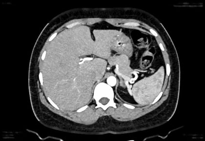 Actual image from Veda
Liver, pancreas, kidneys, bowel and the urinary tract. CT KUB finds a stone in minutes with no preparation at all; the triple-phase study is for characterising a liver lesion.
CT KUB needs nothing — walk in. For every contrast study come after 4 hours of fasting with a recent creatinine report, and bring previous ultrasound or CT films so we can compare.
CT KUB under a minute. A triple-phase study takes about 20 minutes because we scan three times as the dye moves.
Same day.
- CT Abdomen plain
- CT Abdomen contrast
- CT KUB
- CT IVP
- Triple phase CT
- CT Enterography
- CT Fistulogram
Spine
About this group▾
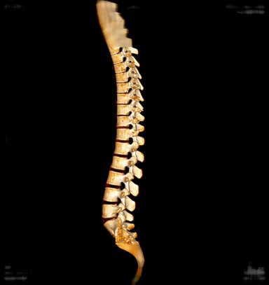 Actual image from Veda
Shows the bone of the spine in far more detail than an X-Ray — fractures, slipped vertebrae, and the exact state of the bone before spinal surgery or after a fall.
No preparation. Remove belts and anything metal from your pockets. If you have had spine surgery, bring the operation notes and the implant details.
Under a minute.
Same day.
- Cervical
- Dorsal
- Lumbar
- Whole spine
Emergency & trauma
About this group▾
After a serious accident the whole body is scanned in one pass — head, neck, chest, abdomen and pelvis together — so that nothing is missed while the patient is still on the table.
None. This runs 24 hours and needs no booking. Bring the patient straight in; we do the paperwork afterwards.
A few minutes from arrival to the last image.
Reported immediately. Dr. Mate is contactable on the doctors' line at any hour.
- Polytrauma whole-body protocol — single pass
Children
About this group▾
Children are not small adults. We use protocols that cut the radiation dose to a fraction of the adult setting while still answering the question the paediatrician asked.
Tell us the child's weight when you book — the dose is set from it. A parent may stay in the room with a lead apron. Bring a toy or a phone with a video to keep the child still.
Under a minute.
Same day.
- Paediatric low-dose protocols
Every scan performed personally by Dr. Shriram B. Mate
Ultrasonography
About this group▾
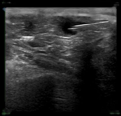 Actual image from Veda
Sound waves, no radiation at all — safe to repeat as often as needed. Covers the abdomen, kidneys, prostate, thyroid, scrotum, soft-tissue lumps and the appendix. Every scan here is done by Dr. Mate himself.
For the abdomen, come after 6 hours of fasting. For the kidneys, bladder or prostate, drink 3 to 4 glasses of water an hour before and do not pass urine. A thyroid, scrotum or lump scan needs no preparation at all.
10 – 20 minutes.
Handed to you immediately after the scan, and explained by Dr. Mate himself.
- Abdomen & pelvis
- Pelvis
- KUB / prostate
- Scrotum
- Chest
- Thyroid / neck
- Neuro-sonography (NSG)
- Musculoskeletal
- Orbit (B-scan)
- Fistulogram
- Appendix — graded compression
- Hernia — dynamic, with Valsalva
- Soft tissue lump and salivary gland
- Transrectal ultrasound (TRUS) prostate, with or without biopsy
- Lung ultrasound
Fetal medicine & women's imaging
About this group▾
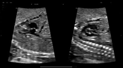 Actual image from Veda
Everything from the first dating scan to the detailed 18–22 week survey of the baby's brain, spine, heart, kidneys, face and limbs — plus growth, fluid, the placenta and cervical length. Dr. Mate is Fetal Medicine Foundation UK certified.
No fasting — eat normally. Bring your antenatal card, the date of your last period, and every previous scan report. Allow extra time: if the baby is lying awkwardly we may ask you to walk for ten minutes and come back. One relative may sit with you.
An anomaly scan takes 30 to 45 minutes. A growth or Doppler scan takes about 20.
Immediately after the scan — Dr. Mate explains the findings himself. Form F is completed as the law requires. We do not reveal the sex of the baby under any circumstances.
- Early obstetric scan
- NT / NB scan
- Level 1 anomaly scan
- Level 2 anomaly scan / TIFFA
- Fetal echocardiography
- Pre-eclampsia risk assessment
- Aneuploidy risk assessment
- 3D / 4D fetal scan
- Obstetric Doppler
- Follicular / ovulation study
- Transvaginal (TVS) / pelvic scan
- Breast / sono-mammography
- Fetal neurosonogram
- Cervical length — preterm birth screening
- Fetal growth and serial growth scan
- Biophysical profile (BPP)
- Twin protocol and TTTS surveillance
- Antral follicle count / ovarian reserve
Colour Doppler
About this group▾
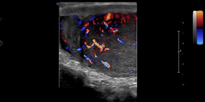 Actual image from Veda
Shows blood moving and measures how fast. For leg swelling and suspected clot, varicose veins, narrowed neck arteries, kidney and liver blood flow, and checking a dialysis fistula before and after surgery.
For a leg, arm or neck study, nothing at all. For kidney or liver Doppler, come after 6 hours of fasting. Wear loose clothing that rolls up easily.
20 – 30 minutes. Both legs take longer than one.
Immediately after the scan.
- Lower limb venous
- Lower limb arterial
- Upper limb venous / arterial
- Renal Doppler
- Carotid Doppler
- Portal Doppler
- Pre & post-operative AV fistula
- Obstetric Doppler
- Carotid intima-media thickness (CIMT)
- Transcranial Doppler
- Penile Doppler
- Varicose vein and pre-bypass vein mapping
- Ocular and orbital Doppler
Elastography & contrast
About this group▾
Two things a standard ultrasound cannot do. Elastography measures how stiff the liver is, which is how fatty liver and fibrosis are staged without a biopsy. Contrast ultrasound characterises a liver lesion using a bubble injection, with no radiation and no CT dye.
Come after 6 hours of fasting — food changes the stiffness reading. Bring your liver function tests and any previous ultrasound or fibroscan report so the numbers can be compared over time.
15 minutes for elastography, 25 for contrast ultrasound.
Immediately after the scan, with the stiffness value and its fibrosis stage.
- Liver elastography — fibrosis and fatty liver staging
- Contrast-enhanced ultrasound (CEUS)
- Shear-wave elastography — thyroid and breast
Children
About this group▾
Ultrasound is the right first test in a child because there is no radiation. Hip screening in a newborn, pyloric stenosis in a vomiting infant, and intussusception — which we can often reduce under ultrasound guidance instead of surgery.
For hip screening, bring the baby fed and settled — a crying baby tenses the hip and the measurement suffers. For a suspected pyloric stenosis, do not feed for two hours before. Bring the paediatrician's note.
10 – 15 minutes.
Immediately after the scan.
- Paediatric hip (Graf) — DDH screening
- Infantile hypertrophic pyloric stenosis
- Intussusception — diagnosis and guided reduction
Allengers digital, with stereotactic biopsy — 24 hours
Mammography
About this group▾
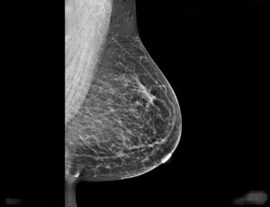 Actual image from Veda
A low-dose X-Ray of the breast, and the only test proven to find cancer before it can be felt. Screening for women with no symptoms, diagnostic views for a lump or discharge, and stereotactic biopsy on the same machine when something needs sampling.
If you still have periods, book in the week after your period ends — the breasts are least tender then. Do not use talcum powder, deodorant, perfume or cream on the chest or underarms that day. Bring every previous mammogram and film: comparison is what makes a small change visible. Wear a two-piece outfit. A female attendant is present throughout.
10 – 15 minutes. The breast is pressed firmly for a few seconds at a time — uncomfortable rather than painful, and quickly over.
Same day, with a BI-RADS category and a clear next step. Available 24 hours.
- Cranio-caudal (CC) views
- Medio-lateral oblique (MLO) views
- Screening mammography
- Diagnostic mammography
- Stereotactic biopsy
- Sono-mammography
- Spot compression and magnification views
- Male mammography / gynaecomastia
Allengers 300 mA high-frequency true digital radiography
Chest
About this group▾
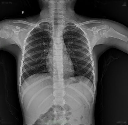 Actual image from Veda
The most-requested X-Ray there is. Shows the lungs, heart size and the ribs — for cough, fever, breathlessness, chest pain, and before surgery or a job medical.
Nothing. Remove chains, lockets and anything in your shirt pocket. You stand against the plate, take a deep breath and hold it. Tell the technician before the exposure if you may be pregnant.
Two minutes.
Same visit, in most cases. Digital, so the image is on screen before you have left the room.
- Chest
- Sternum
- S.C. joints
- Lateral, apical lordotic and decubitus views
Abdomen
About this group▾
For sudden severe abdominal pain, vomiting and constipation together, or a swallowed object. The erect and supine pair together show obstruction and free air under the diaphragm.
None for an emergency — walk straight in, this runs 24 hours. Empty your bladder first if you can. Remove belts and trouser studs.
Three to five minutes for the pair.
Same visit. Emergencies reported immediately.
- Single view (KUB)
- Acute abdomen (includes chest PA)
- Erect and supine abdomen series
Head & neck
About this group▾
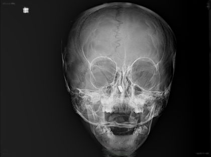 Actual image from Veda
Skull, sinuses, nasal bones, jaw and the soft tissue of the neck. Used after a facial injury, for a blocked nose, and in a child with noisy breathing or a swallowed foreign body.
None. Remove earrings, nose pins, spectacles, hairpins and any removable denture. For sinus views you will be asked to sit upright with your chin raised.
Three to five minutes.
Same visit.
- Skull
- PNS
- Nasal bones
- Mandible
- T.M. joints
- Orbits
- Neck
- Soft tissue neck — adenoids and foreign body
Upper extremities
About this group▾
 Actual image from Veda
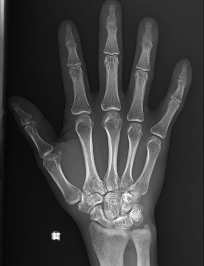
Shoulder, collarbone, arm, elbow, forearm, wrist and hand. Mostly after a fall or an injury, and for arthritis. The scaphoid view is taken separately because that small wrist bone hides on an ordinary film.
None. Remove rings, bangles, watch and any plaster if your doctor has said it can come off — otherwise we scan through it. Tell us which side hurts; we always take the other side too if a comparison helps.
Two to five minutes.
Same visit.
- Shoulder
- Clavicle
- A.C. joints
- Scapula
- Humerus
- Elbow
- Forearm
- Wrist
- Scaphoid
- Hand
Lower extremities
About this group▾
 Actual image from Veda
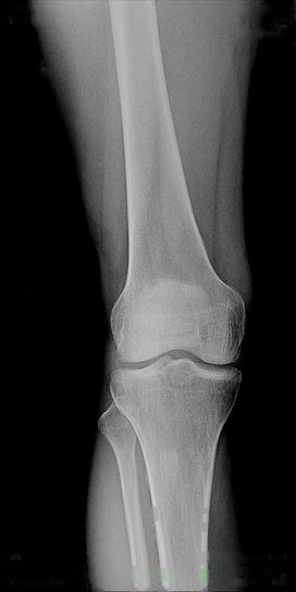
Thigh, knee, leg, ankle, foot and heel. The weight-bearing knee film is taken standing, not lying down — that is how orthopaedic surgeons grade arthritis, and a supine film will usually be sent back.
None. Wear something that rolls up above the knee, or a loose skirt. For the standing knee film you will be asked to put equal weight on both legs — tell us if that is painful and we will support you.
Three to five minutes. A full-length standing lower limb film takes about ten.
Same visit.
- Femur
- Knee
- Tib & fib
- Ankle
- Foot
- Calcaneus
- Weight-bearing knee
- Full-length standing lower limb — hip-knee-ankle alignment
Spine & pelvis
About this group▾
 Actual image from Veda
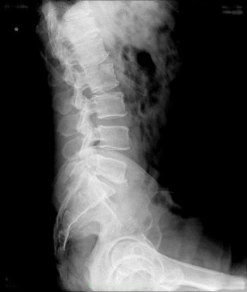
Neck, mid-back, lower back, sacrum, the sacroiliac joints and the hips. Flexion and extension views are taken bending forward and back, to show whether a vertebra slips when you move.
For the lower back the bowel should ideally be empty — your doctor may advise a laxative the night before. Remove belts, trouser studs and anything in your pockets. Tell us before the film if you may be pregnant.
Five minutes. Flexion and extension views take about ten.
Same visit.
- Cervical spine
- Thoracic spine
- LS spine
- Sacrum & coccyx
- S.I. joints
- Pelvis & hip (PBH)
- Scoliosis series
- Cervical flexion and extension
- Lumbar flexion and extension
Skeletal survey
About this group▾
A fixed set of films covering the whole skeleton, taken to one protocol so nothing is missed. Used to look for spread of cancer into bone, in myeloma, in metabolic bone disease, and to assess bone age in a child.
Allow a longer appointment — this is many films, not one. Bring every previous film and report, and your blood reports if you have them. Wear something simple that is easy to change out of.
30 – 45 minutes.
Same day.
- Metastatic series
- Arthritic series
- Metabolic series
- Bone age
- Skeletal survey for suspected non-accidental injury
Other
About this group▾
The studies that do not fit a body part — locating a swallowed or embedded foreign body, checking a plate or screw after surgery, stress views that are taken while the joint is pushed, and the scanogram for limb length.
Tell us exactly what is being looked for — a foreign body film is taken differently depending on whether the object is metal, glass or bone. For post-operative films, bring the operation notes and the implant details.
Five minutes.
Same visit.
- Scannogram
- Foreign body localisation
- Post-operative implant and fracture follow-up
- Stress views — ankle and AC joint
Fitness & certification
About this group▾
A chest X-Ray with a signed radiologist's report, for a job, a visa or a medical fitness certificate. The report carries our registration details and Dr. Mate's signature, which is what the receiving office asks for.
Bring a photo ID and the form your employer or consulate has given you, if there is one — some want the report worded a particular way, and it is far easier to do that at the time than afterwards.
Ten minutes including the paperwork.
Same visit, signed and stamped.
- Pre-employment and visa chest X-Ray
Brand-new Vatech digital OPG
OPG
About this group▾
 Actual image from Veda
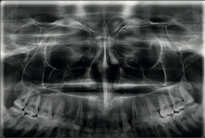
One wide picture showing every upper and lower tooth, both jaws and both jaw joints together. Standard before an extraction, before braces, and before an implant. Periapical and occlusal views cover a single tooth in close detail.
Remove earrings, nose pin, chains, spectacles, hairpins and any removable denture. Nothing else — no fasting. Carry your dentist's referral slip.
Under a minute. You stand still and the machine moves around your head.
Film and report within 15 minutes.
- Orthopantomogram (OPG)
- Full pano examination
- Bitewing
- TMJ open / close
- Sinus PA
- Lateral cephalogram
- Periapical and occlusal views
CBCT
About this group▾
A true three-dimensional scan of the jaw. It shows bone height, bone width and exactly where the nerve runs — which is what an implant cannot be planned without. Also for impacted teeth, root canal assessment, the jaw joint, the sinuses and facial trauma.
No fasting. Remove all metal from the head and neck. Bring your dentist's referral slip — it tells us which region and how large a field of view to capture, and taking the wrong one means repeating the scan.
Two to three minutes. The dose is far lower than a medical CT of the head.
Films the same visit, plus a CD of DICOM data that opens in standard implant planning software. We can also export an STL file for a surgical guide.
- CBCT single tooth or region
- CBCT for implant planning
- CBCT for impacted teeth
- CBCT before and after root canal
- TMJ imaging
- Maxillary sinus and airway assessment
- CBCT airway analysis for sleep apnoea
- Canine and supernumerary tooth localisation
- CBCT for maxillofacial trauma
- Paranasal sinus CBCT — low dose
- STL / DICOM export for surgical guides and 3D printing
Orthodontic
About this group▾
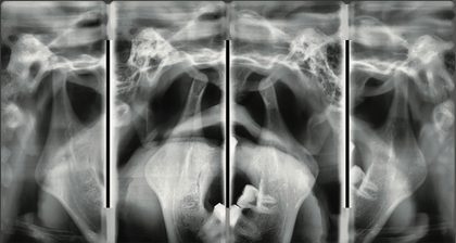 Actual image from Veda
The films an orthodontist needs before starting treatment — cephalograms to measure jaw relationships, 3D cephalometric analysis from the CBCT, and a hand-wrist film that tells you how much growth is left.
No preparation. Remove all metal from the head. For a hand-wrist film, bring the child's date of birth — the reading is compared against it. Carry the orthodontist's tracing sheet if they have given you one.
Under a minute each.
Same visit, with the tracing measurements where they have been requested.
- Hand-wrist film for skeletal maturity
- 3D cephalometric analysis from CBCT
Performed personally by Dr. Shriram B. Mate
Ultrasound-guided
About this group▾
Actual image from VedaA needle placed while the ultrasound is running, so it reaches the right spot without an operation. Biopsy and FNAC of liver, kidney, breast, thyroid and lymph node; draining fluid from the chest or abdomen; abscess drainage; nephrostomy; and cyst aspiration.
Come after 6 hours of fasting. A recent PT/INR and platelet count is required before any biopsy or drainage. Tell us if you take aspirin, clopidogrel, warfarin or any blood thinner — these usually have to be stopped first, but only on your treating doctor's advice, never on your own. Bring a relative; you should not drive yourself home.
20 – 45 minutes, then a short period of observation here before you go home. Local anaesthetic is given first, so you feel pressure rather than pain.
The imaging report the same day. Biopsy and FNAC samples go to the pathology lab of your choice, and that report comes on the lab's own timeline — usually two to four days.
- Pleural aspiration / tapping
- Ascitic aspiration / tapping
- Liver abscess drainage
- Any other drainage
- Liver / renal / breast biopsy
- FNAC
- Percutaneous nephrostomy (PCN)
- Pigtail catheter and intercostal drain
- Thyroid and lymph node FNAC
- Cyst aspiration and sclerotherapy
CT-guided
About this group▾
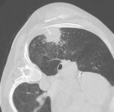 Actual image from Veda
When the target is deep, behind bone or inside the lung, CT guidance places the needle where ultrasound cannot see. Lung and bone biopsy, cisternography, dacryocystography, and the CT versions of colonoscopy and bronchoscopy.
The same fasting and blood-thinner rules as an ultrasound-guided procedure, plus a recent chest film and lung function report for a lung biopsy. Written consent is taken and the whole procedure is explained to you before anything begins.
30 – 45 minutes, followed by a longer period of observation after a lung biopsy.
The imaging report the same day; the pathology report on the lab's timeline.
- CT-guided lung biopsy
- CT bone biopsy
- CT distal loop cologram
- CT colonoscopy
- CT bronchoscopy
- Dynamic contrast esophagogram
- CT cisternography
- CT dacryocystography (DCG)
X-Ray procedures
About this group▾
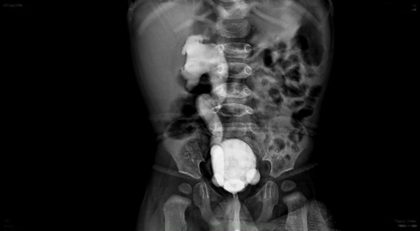 Actual image from Veda
Contrast studies done under live X-Ray — HSG for the fallopian tubes, MCU and urethrography for the bladder and urethra, IVP for the kidneys, barium studies for the food pipe, stomach and bowel, and fistulography.
Barium studies need overnight fasting, so book a day ahead. HSG is done between day 7 and day 11 of your cycle, after bleeding has stopped and before ovulation — and you must not be pregnant. A female attendant is present throughout. Bring your referring doctor's note.
20 – 40 minutes depending on the study. A barium follow-through takes longer because films are taken as the barium moves.
Same day.
- HSG
- IVP
- ASU / RGU
- MCU
- DCG
- Barium swallow, meal, follow-through and enema
- Scannogram
- Fistulogram — sinogram
Musculoskeletal intervention
About this group▾
Actual image from VedaInjections placed under live ultrasound, so the drug goes exactly where it is needed instead of near it. Joint injection and aspiration, hydrodilatation for a frozen shoulder, barbotage for calcific tendinitis, PRP, plantar fascia and tendon sheath injections, and nerve root, facet joint and epidural blocks.
No fasting for most injections. Bring your MRI or X-Ray films — the injection is planned from them. Tell us about any blood thinner, and about diabetes, because a steroid injection can raise your sugar for a day or two. Arrange for someone to drive you home after a spine injection.
15 – 30 minutes. Most people walk out and go home the same hour.
A note of what was injected and where, the same day, for your orthopaedic surgeon or pain physician.
- Joint injection and aspiration
- Hydrodilatation for frozen shoulder
- Barbotage for calcific tendinitis
- PRP and regenerative injection
- Plantar fascia and tendon sheath injection
- Nerve root, facet joint and epidural injection
Any other customised scan as per your request.
Print this page for your desk, or send the link to your patient. Dr. Mate is available on the doctors' line for case discussion before or after any study.